
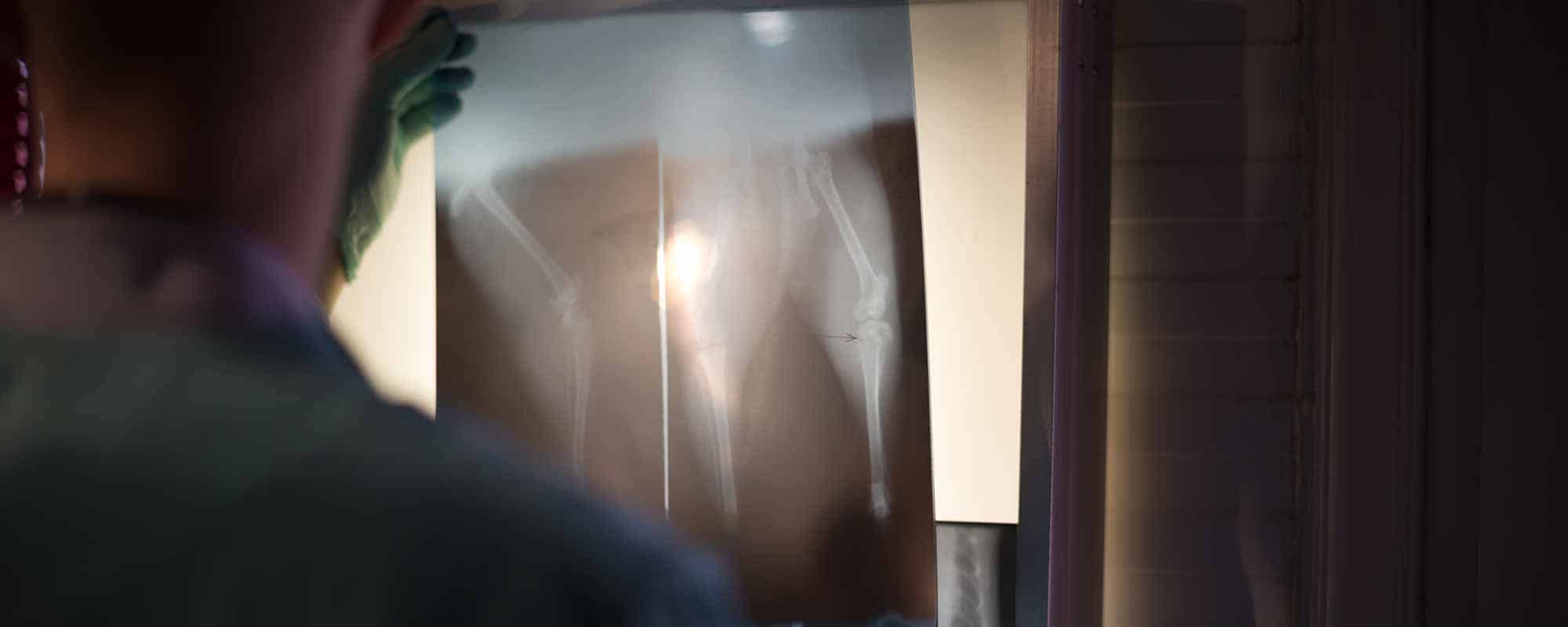
Advanced Veterinary Diagnostic Imaging in Southeastern Michigan
When a pet is sick or injured, the answers are not always visible from the outside. Diagnostic imaging lets veterinary teams look beneath the surface, evaluate internal structures, and gather information to guide the next steps in care.
Oakland Veterinary Referral Services offers advanced imaging for dogs, cats, and other companion animals with complex medical needs. Our Diagnostic Imaging team works closely with OVRS specialists and referring veterinarians to help identify disease, evaluate injuries, plan treatment, and monitor a patient’s progress.
Whether your pet needs an ultrasound, CT scan, digital radiographs, or another specialized study, our goal is to obtain the information your veterinary team needs while keeping your pet as safe and comfortable as possible.
Seeing More Clearly—and Treating More Precisely
Diagnostic imaging can help veterinarians evaluate the size, shape, location, and function of structures inside the body without exploratory surgery.
Imaging may be recommended to:
- Investigate unexplained symptoms
- Evaluate an injury
- Examine an organ or body system in greater detail
- Identify the location and extent of a mass
- Look for internal bleeding or fluid
- Evaluate bones, joints, the spine, or soft tissues
- Determine whether a disease has spread
- Plan surgery or another procedure
- Guide treatment decisions
- Monitor a known condition or response to treatment
Different imaging methods reveal different types of information. Your veterinarian or OVRS specialist will select the study—or combination of studies—most likely to answer the clinical questions affecting your pet.
Diagnostic Imaging Services at OVRS
Digital Radiography
Digital radiography produces X-ray images of bones and internal structures. Images are available quickly and can be adjusted electronically to help the veterinary team evaluate areas of concern in greater detail.
Radiographs may be used to examine:
- Bones and joints
- The chest and lungs
- The abdomen
- The spine
- Dental or facial structures
- Certain foreign objects
- Changes in organ size or position
Digital radiographs are frequently used as an initial diagnostic tool and may reveal whether additional imaging is needed.
The procedure itself is not painful, although a sore or injured pet may be uncomfortable when positioned. Some patients can remain awake, while others may benefit from sedation for comfort, safety, or accurate positioning.
Veterinary Ultrasound
Ultrasound uses sound waves to create real-time images of structures within the body. It does not use ionizing radiation and can show the appearance, movement, and internal architecture of many organs and soft tissues.
Ultrasound may be used to evaluate:
- Liver and gallbladder
- Kidneys and adrenal glands
- Bladder and urinary tract
- Stomach and intestines
- Pancreas
- Spleen
- Lymph nodes
- Prostate and reproductive organs
- Abdominal fluid
- Masses or other tissue abnormalities
Because ultrasound displays moving images, the veterinary team can observe organ motion, blood flow in selected studies, and changes that may not be apparent on a still radiograph.
Many pets do not require sedation or anesthesia for an ultrasound. However, medication may be recommended if a patient is painful, anxious, unable to remain still, or undergoing an image-guided procedure.
A section of hair usually must be clipped so the ultrasound probe can make proper contact with the skin. The technician applies a water-based gel to the area during the examination.
Computed Tomography—CT
Computed tomography, commonly called a CT scan, uses X-rays and computer processing to produce detailed cross-sectional images of the body. These images can be viewed individually or reconstructed to provide a three-dimensional understanding of complex anatomy.
CT is particularly useful for examining areas that may be difficult to evaluate fully with traditional radiographs, including:
- Brain and skull
- Nasal passages and sinuses
- Inner and middle ears
- Eye sockets and surrounding tissues
- Spine and intervertebral discs
- Bones and joints
- Chest and lungs
- Soft-tissue masses
- Complex fractures
- Areas being evaluated for surgery or radiation treatment
Contrast material may be administered in some studies to make blood vessels, organs, or abnormal tissues easier to distinguish.
Because movement can blur CT images, patients generally require sedation or anesthesia to remain safely and precisely positioned. The team will develop an individualized plan based on your pet’s health and the type of study being performed.
Fluoroscopy and Contrast Studies
Fluoroscopy uses a continuous or pulsed X-ray beam to create moving images. While conventional radiographs capture individual still images, fluoroscopy allows the veterinary team to observe motion and function in real time.
It may be used to evaluate:
- Swallowing disorders
- Movement through the esophagus
- Selected gastrointestinal problems
- Airway function
- Joint movement or instability
- Certain interventional or surgical procedures
A contrast substance may be given to outline structures that would otherwise be difficult to see.
Specialized contrast studies available through OVRS may include upper gastrointestinal examinations and portograms. A portogram evaluates blood flow through the liver and may help diagnose or plan treatment for a portosystemic shunt.
Preparation, sedation, and duration vary. Your veterinary team will explain what your pet’s particular procedure involves.
Echocardiography
Echocardiography is a specialized ultrasound examination of the heart. It provides detailed, moving images that allow veterinary cardiologists to evaluate the heart’s structures and function.
An echocardiogram may be recommended for pets with:
- A heart murmur
- Suspected heart disease
- Abnormal heart rhythm
- Fluid around the heart or lungs
- Exercise intolerance or fainting
- A suspected mass involving the heart
- A need for pre-anesthetic cardiac evaluation
- A heart condition requiring continued monitoring
At OVRS, the Cardiology Department performs and interprets echocardiography. Learn more about Veterinary Cardiology at OVRS.
Advanced Imaging for Surgical and Treatment Planning
Imaging does more than establish a diagnosis. It may also provide a detailed roadmap for surgery or another treatment.
CT and other imaging techniques can help a specialist:
- Determine the precise location and extent of a lesion
- Understand its relationship to nearby organs, bones, nerves, or blood vessels
- Evaluate complex fractures or joint abnormalities
- Assess whether a mass may be surgically accessible
- Select an appropriate surgical approach
- Prepare for reconstructive or minimally invasive procedures
- Compare findings before and after treatment
Because OVRS houses multiple specialties within one hospital, radiologists and imaging professionals can communicate directly with surgeons, internists, oncologists, emergency clinicians, cardiologists, and other specialists involved in a patient’s care.
What to Expect When Your Pet Needs Imaging
The details of your pet’s visit will depend on the imaging study and the medical reason for the request.
Before the appointment, the team may ask you to:
- Withhold food for a specified period
- Allow continued access to water
- Give or temporarily withhold certain medications
- Bring previous images or arrange for them to be transferred
- Provide recent laboratory results and medical records
- Arrive at a particular time for admission or preparation
Follow the instructions provided specifically for your pet. Do not withhold food, water, or medication unless your veterinarian or the OVRS team directs you to do so.
At the hospital, the team will review your pet’s history and the clinical questions the imaging study is intended to answer. Depending on the procedure, your pet may need:
- A small area of hair clipped
- An intravenous catheter
- Sedation or anesthesia
- Contrast material
- Monitoring before, during, and after the study
- A brief recovery period before going home
The team will explain any special preparation, anticipated risks, expected timing, and discharge instructions.
Will My Pet Need Sedation or Anesthesia?
Not every imaging procedure requires sedation or anesthesia.
The decision depends on:
- The type of study
- The position your pet must maintain
- How long the procedure takes
- Your pet’s comfort and anxiety level
- The presence of pain or injury
- The need for precise, motion-free images
- Your pet’s overall health
Many ultrasound examinations can be performed while a pet is awake. Radiographs sometimes require sedation for positioning, while CT generally requires sedation or anesthesia to prevent movement.
When medication is needed, the veterinary team develops an individualized plan and closely monitors your pet.
What Happens After the Images Are Taken?
Producing an image is only one part of diagnostic imaging. The images must also be evaluated in the context of your pet’s symptoms, examination findings, laboratory results, and medical history.
After review, we communicate imaging findings to the veterinarian or specialist managing your pet’s care. That clinician will discuss what the results mean, whether additional testing is needed, and how the findings may affect treatment.
In urgent hospitalized cases, imaging results may guide immediate care. For scheduled or externally submitted studies, reporting time depends on the service type, case complexity, and current availability.
Collaborative Specialty Care at OVRS
A single patient may need expertise from several departments.
For example:
- A surgeon may use CT images to plan a complex procedure.
- An oncologist may use imaging to assess a tumor and look for evidence of spread.
- An internist may request ultrasound to investigate changes in the liver, kidneys, pancreas, or gastrointestinal tract.
- An emergency clinician may use imaging to look for internal injury or bleeding.
- A cardiologist may perform an echocardiogram to evaluate heart structure and function.
Advanced imaging and multiple specialties within the same hospital support timely communication and a more coordinated approach to complex cases.
Partnering With Your Family Veterinarian
Your family veterinarian remains an important part of your pet’s healthcare team.
OVRS communicates relevant imaging findings and recommendations to the referring veterinarian and other specialists involved in the case. Depending on the results, follow-up care may take place at OVRS, with your family veterinarian, or through a coordinated combination of both.
This partnership helps ensure that imaging findings translate into a practical treatment plan for your pet.
Preparing for Your Pet’s Visit
Before arriving:
- Follow all fasting and medication instructions.
- Ask your veterinarian to send pertinent medical records.
- Arrange for previous radiographs, ultrasound images, CT studies, and reports to be transferred.
- Bring a current list of medications and supplements.
- Let the team know about previous reactions to sedation, anesthesia, or contrast material.
- Tell us if your pet’s condition changes before the appointment.
- Bring your questions about the procedure and expected next steps.
If you are unsure about any instructions, please call before the visit.
Advanced Images. More Informed Decisions.
Diagnostic imaging helps turn uncertainty into useful information. By seeing internal structures more clearly, veterinary teams can diagnose disease, plan treatment, and make more informed decisions about a pet’s care.
At OVRS, advanced technology is supported by experienced professionals, collaborative specialty care, and a commitment to treating every patient with compassion.
Ask your family veterinarian whether advanced diagnostic imaging may be appropriate for your pet, call OVRS at (248) 334‑6877, or request an appointment.
Information for Referring Veterinarians
The OVRS Diagnostic Imaging Department supports referring veterinarians with advanced imaging and, when available, external image-interpretation services.
Services may include:
- Review of submitted radiographic studies
- Collaboration on challenging imaging cases
- Digital radiography
- Ultrasound
- CT
- Fluoroscopy and contrast studies
- Advanced imaging for surgical planning
Availability, submission procedures, reporting times, and fees may change. Please contact radiology@ovrs.com for current information about external image interpretation or special circumstances.