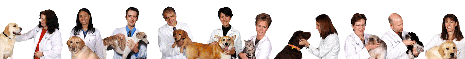
Veterinary Radiology: From Diagnosis to Treatment
Out of all the advancements in veterinary medicine, almost none is as important as radiology. Veterinary diagnostics give us a clearer picture of what is happening inside your pet so we can make the most informed treatment decision. Keep reading to see how veterinary radiology is an invaluable tool from the first moment we see your pet until we successfully treat them:
What Is Veterinary Radiology?
Veterinary radiology is a term that encompasses the diagnostics and imaging we use to build a complete picture of your pet’s health. This includes:
- Digital radiography
- Ultrasound
- Echocardiology
- Computer Tomography (CT) Scan
- Upper gastrointestinal imaging
- Fluoroscopy
- Portogram
- Advanced imaging for surgery planning
We determine which forms of radiology to use based on a pet’s issues and symptoms so we can offer the best treatment for his or her needs.
Veterinary Digital Radiography
Whether your dog is dealing with a broken bone, vomiting, or a concern for a foreign body or mass, the vet might take a radiograph. This uses electromagnetic energy to take pictures inside your pet’s body. The X-rays are either scattered, absorbed, or penetrate the tissues depending on the characteristics of the tissue to create the image.
It usually only takes about 10 minutes to take the pictures. The procedure may require temporary sedation for adequate positioning to obtain diagnostic images. Digital radiography creates images instantly, so your veterinarian has immediate access to the pictures to move forward with decisions about treatment.
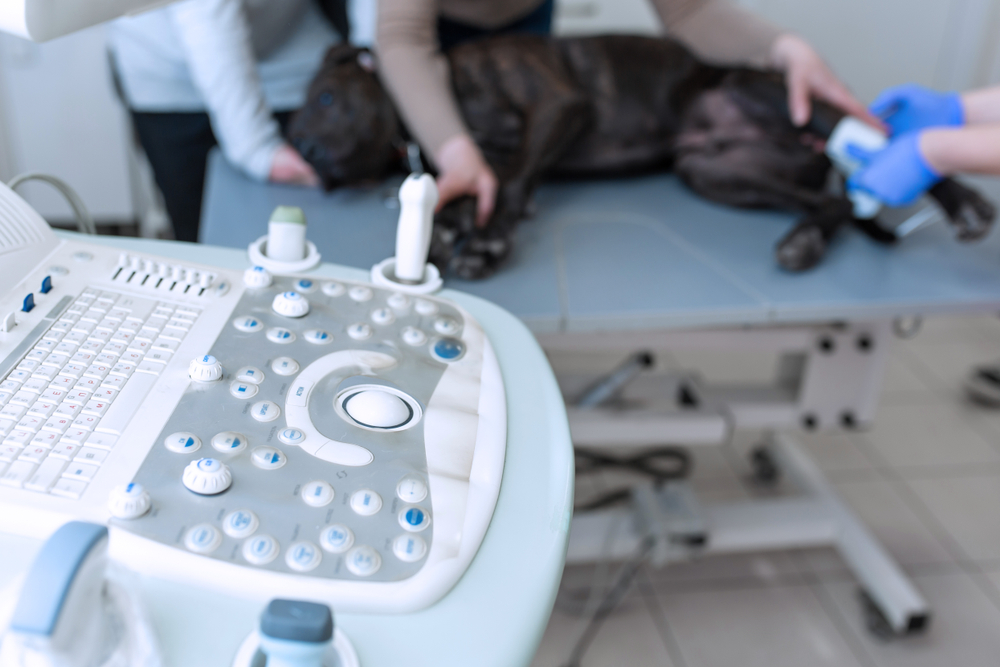
Veterinary Ultrasound
Veterinary ultrasound uses non-invasive sound waves to create interior images of your pet’s body. Ultrasound is often a complementary imaging modality to X-rays and may be recommended in addition to radiographs of your pet if there is a specific concern to evaluate.
Veterinary ultrasound in our facility is performed and interpreted by a boarded specialist. This procedure is non-invasive and often relaxing for your pet, with them positioned on their side or back. Like radiographs, it may require light or temporary sedation to obtain diagnostic images. It is extremely helpful in identifying and diagnosing potential problems early as well as localizing a problem that may have been identified on radiographs. Using ultrasound with the pet under light sedation, we may recommend obtaining an ultrasound-guided needle sample of an organ to evaluate by a pathologist under a microscope (called cytology). Ultrasound can be used to evaluate:
- The liver
- The gallbladder and biliary tract
- The kidneys
- The adrenal glands
- The urinary bladder
- The stomach
- The small intestine
- The pancreas
- The colon
- The peritoneum
- The spleen
- The lymph nodes
- The prostate
- The uterus and ovaries
Veterinary CT Scans
CT scanners are noninvasive diagnostic tools that allow our specialists to view your pet’s internal anatomy by producing highly detailed cross-sectional images using digital X-rays and a computer. These two-dimensional slices it captures are reconfigured into a complete image. Scans provide excellent detail of bony and soft tissue structures in the body.
CT scans are often requested as part of diagnosing disease, planning surgery, and early detection and treatment of cancer. CT scans are commonly requested in diagnosing disease that affects these areas:
- Spine
- Nasal cavity/Sinuses
- Brain
- Chest and Lungs
- Inner ears
- Orbit
- Intervertebral disc
- Bones and Joints
- Soft tissues
- Lymph nodes
- Thyroid
- Abdominal organs
- Vascular structure
A contrast agent given intravenously (IV) allows us to see blood flow in the body, aiding in cancer detection. We can time the CT scanner in relation to the timing of the IV contrast to better visualize structures of concern and to determine the arteries and vein positioning and relation to masses, clots within the vessels, or the patterns of contrast enhancement within these structures.
The CT scans in our hospital are overseen and interpreted by our radiologists, with the protocols for the CT scan individually tailored to each patient’s needs. Traditional fan-beam CT scans are recommended as opposed to cone-beam CT scans when evaluating all anatomy except for dental structures.
Unfortunately, we can’t simply ask pets to lie still and hold their breath during a CT scan, so this tool requires a brief period under anesthesia or heavy sedation.
Echocardiography
This diagnostic imaging tool is critical for our cardiology team. It provides incredibly detailed views of the heart’s function, helping to diagnose heart disease and monitor ongoing treatment. Echocardiography uses ultrasound waves to see how well the heart is functioning, how well the valves are performing, and any detectable defects. Echocardiogram in our hospital is performed and interpreted by our cardiologists.
At Oakland Veterinary Referral Services, we use the most modern tools in veterinary radiology to give your pets access to the best care. From digital radiography to CT scans and others we haven’t discussed, we use these tools to diagnose and treat pets as quickly and as effectively as possible. If your pet is undergoing an imaging procedure, it is important to check that the procedure will be interpreted by a specialist. Ideally, the ultrasound procedure would also be performed by a specialist. OVRS is proud to offer specialty-performed ultrasound examinations and specialist-interpreted imaging procedures, the highest standard of care for patients.
To learn more about our services or to schedule an appointment, please call ( 248) 334-6877.