Diagnostic Imaging for Pets: A Path to Faster, Better Diagnosis
 When your pet is sick, getting a diagnosis is an essential part of getting them well again. The faster an accurate diagnosis is achieved, the faster an accurate, effective treatment plan can be formulated.
Arriving at a diagnosis can involve various types of tests such as blood and fecal testing, but imaging is a cornerstone of diagnostic testing. Oakland Veterinary Referral Services offers a range of diagnostic imaging tools and technology for helping us provide you and your pet with a fast, accurate diagnosis and treatment plan:
-
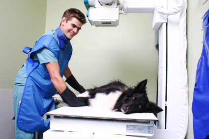
Digital radiography
Radiographs, or “x-rays”, allow our veterinarians a first look at bones and skeletal structure as well as organ shapes, sizes, and characteristics within the abdominal and chest cavities.
Because radiographs show all internal structures as varying shades of gray and only in two dimensions, additional testing may be needed to further investigate abnormalities found on x-rays. Digital x-rays are superior to traditional plain film x-rays as they allow us to produce more detailed images more quickly than in the past. They also make it easy to send the images instantaneously anywhere in the world!
-
Ultrasound
Ultrasound utilizes sound waves to produce visuals of organs and structures within the body in a safe and non-invasive manner. Ultrasound allows the veterinarian to see the characteristics of the organs in more detail than seen on an x-ray, and to observe body processes in real-time. Ultrasound sometimes allows biopsy samples to be taken without the need for actual surgery.
-
Echocardiography
ECGs are an ultrasound of the heart. Because ultrasound lets the veterinarian visualize the heart beating and blood flowing within the chambers, an ECG is an important means to diagnose the cause of heart murmurs, abnormal rhythms, and other cardiac problems.
-
Computed Tomography (CT) Scan
CT scans use the same technology as radiographs to visualize the body in three dimensional “slices”, making it a powerful tool to diagnose problems within the skull and spinal cord among other things.
-
Magnetic Resonance Imaging (MRI)
MRI uses a magnetic field and pulses of radio wave energy to create detailed pictures of organs and other structures within the body. The patient is typically anesthetized for a short period to allow these images to be obtained. MRI is particularly good at creating images of the soft-tissue and neurological structures within the body.
With the aid of these in-house diagnostic imaging tools, we can gain a range of information about what is causing your pet’s injury, illness or pain. We are able to get accurate, detailed information quickly and efficiently. This means we are able to start the right treatment for your pet and get them feeling better again as soon as possible!